UrbanEyes Optometry is now I See Optometry (19 Clarence Street)
Technology
Our clinic proudly uses the best diagnostic equipment.
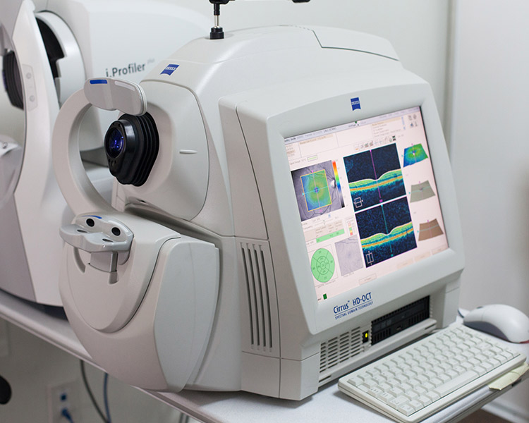
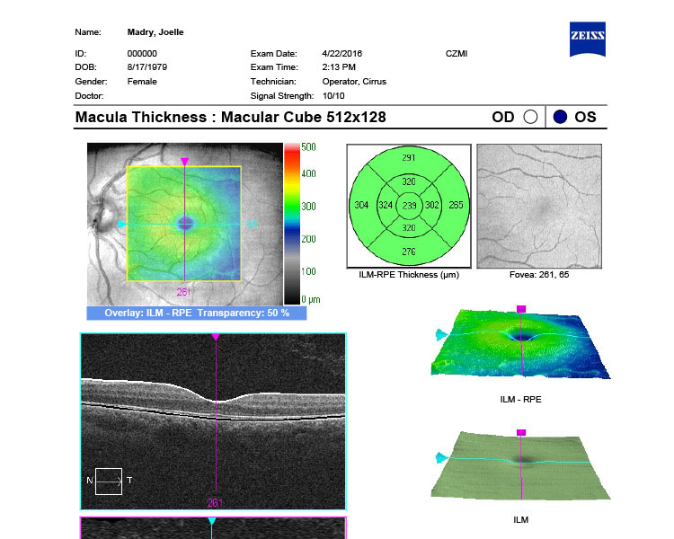
Zeiss Optical coherence tomography
An optical coherence tomography is like a CAT scan of your eye. It is cutting-edge technology that allows our Ottawa optometrists to see structures with a resolution of microns. It uses no radiation, therefore no harm to the patient. It allows our optometrists to visualize microscopic structures such as the very important retina and optic nerves to diagnose glaucoma, macular degeneration and more.
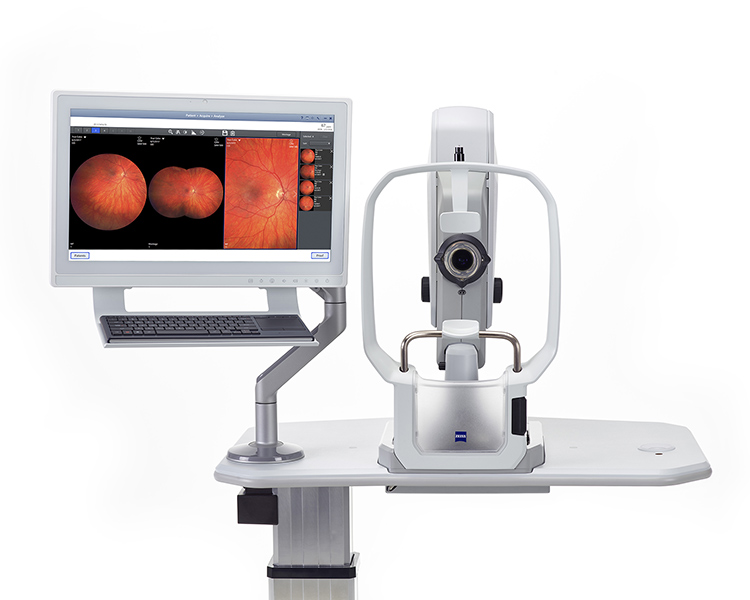
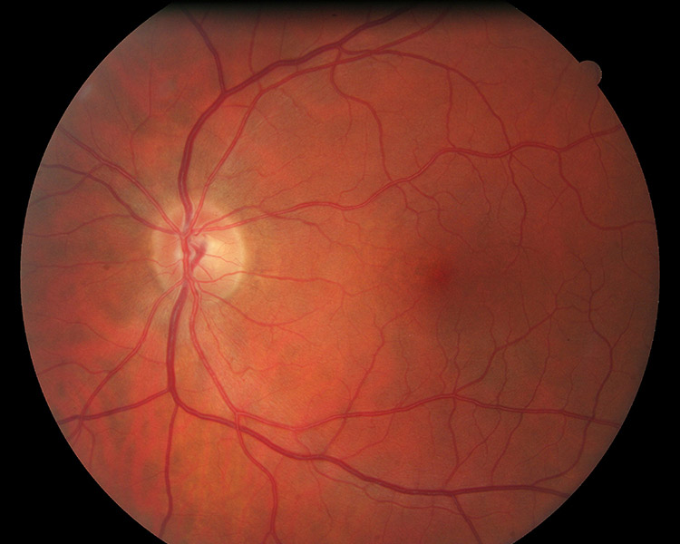
Clarus Digital imaging retinal camera
This advanced retinal camera allows our optometrists to take pictures of the inside of your eye. Every eye has a unique pattern, much like a fingerprint. During your complete eye exam, you will be shown these remarkable photos.
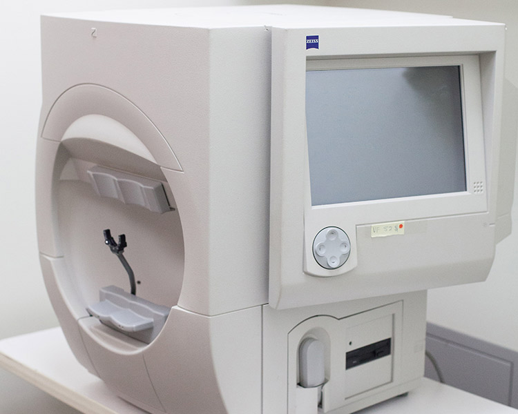
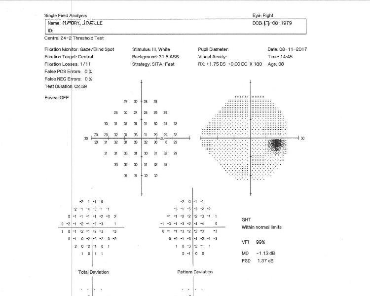
Zeiss Visual field analyzer
This state of the art visual field analyzer allows our optometrists to measure your side vision very precisely. This is critical in management and diagnoses of glaucoma, neurological disorders (such as brain tumors) and genetic retinal degenerations.
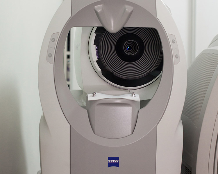
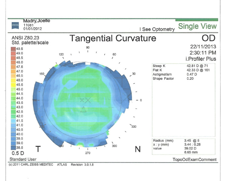
Zeiss i.Profiler®plus corneal & ocular wavefront aberrometer
This innovative tool measures your individual eye’s optics, using a high resolution sensor. In just seconds, the iProfiler maps thousands of points on the corneal surface of the eye! Corneal analysis provides important information, above and beyond an eyeglass prescription, that can help to optimize the quality of your vision. These measurements are critical for refractive surgery, and for the detection and management of corneal pathology, such as keratoconus.
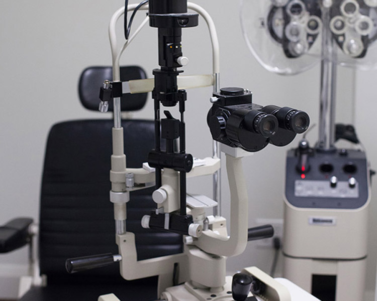
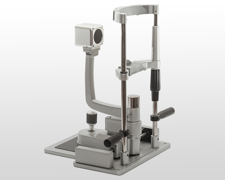
Haag-Streit Biomicroscope
A biomicroscope allows our optometrists to see in great magnified details the important structures of your eye. A Haag-Streit biomicroscope is considered the best one on the market!
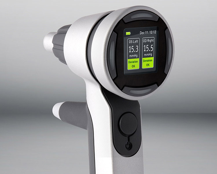
Icare® PRO tonometer
We were the first clinic in Ottawa to use this leading edge measurement of intra-ocular pressure. It does not require the uncomfortable puff of air or numbing drops. Intra-ocular pressure measurements are important for the diagnosis and management of glaucoma.
Meibo
Meibomian Gland Dysfunction is the main etiology of dry eyes. Meibography provides doctors and patients an opportunity to detect and track dry eyes before it is too late. Meibox software allows for the assessment of both upper and lower lids meibomian glands. It can capture 4 images within 4 seconds, it allows the doctor to track the state or progression of the Meibomian Gland Dysfunction.
We are happy to welcome new patients!
| Monday | 8am – 5:30pm |
|---|---|
| Tuesday | 10am – 5:30pm |
| Wednesday | 8am – 8pm |
| Thursday | 8am – 8pm |
| Friday | 9am – 5pm |
| Saturday | 9am – 5pm |
| Sunday | Closed |

